Animals and Preparations
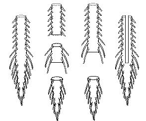
Experiments are performed on isolated spinal cord or brainstem-spinal cord preparations of neonatal rodents with or
without attached hindlimb and tail (for details, see
Lev-Tov et al., 2000,
Delvolve et al., 2001,
Strauss and Lev-Tov, 2003,
Blivis et al., 2007,
Etlin et al. 2010).
We also use the embryonic chick spinal cord for studies of genetically targeted populations
of spinal interneurons (Etlin et al. 2011, Hadas et al. 2011).
Electrophysiological recordings
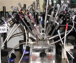
Population recordings from ventral root efferents in the thoracolumbar and sacrocaudal segments are used
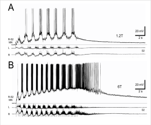
to study the motor output of the preparations. Intracellular recordings are used to determine the physiological
properties and synaptic connectivity of individual spinal neurons in various regions of the spinal cord.
(Lev-Tov et al., 2000,
Strauss and Lev-Tov, 2003,
Blivis et al., 2007).
Visualization of spinal neurons
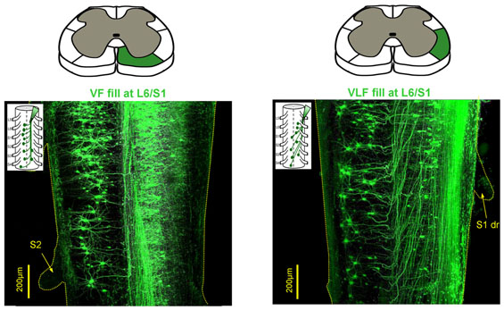
Confocal microscopy imaging of defined populations of fluorescently labeled spinal
neurons and the immuno-labeled contacts between them and other constituents of spinal
networks are used to determine their morphology, axonal projections, spatial distribution and connectomes
(Lev-Tov et al. 2010,
Etlin et al. 2010).
Imaging the activity of spinal neurons during motor behavior
The activity in defined populations of spinal interneurons labeled with
synthetic or genetically encoded calcium sensors is characterized during various
motor behaviors using fluorescence or 2-photon microscopy imaging. The correlation
between the interneuron activity and the simultaneously recorded motor output is examined
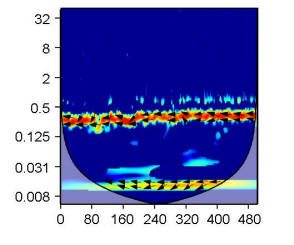
using the Wavelet transformation algorithms (Etlin et al. 2011, Hadas et al. 2011).

Rhythmic activity
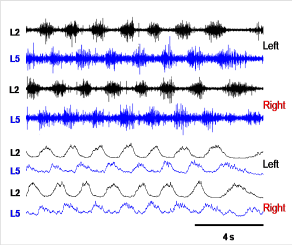
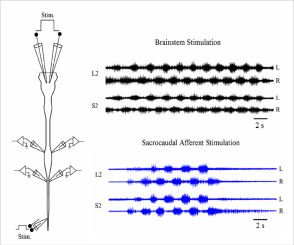
Rhythmic activity is induced by stimulation of
sacrocaudal afferent pathways or by bipolar stimulation of the ventromedial medulla (Blivis
et al., 2007). Neurochemically induced locomotor like
rhythm is initiated by bath-applied NMDA/5HT with or without Dopamine (DA).
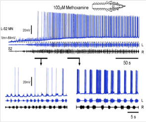
The sacrocaudal rhythm is selectively produced by bath application of the alpha1-adrenoceptor
agonist methoxamine (Gabbay
and Lev-Tov, 2004, Cherniak et al. 2011). When required, the experimental bath
is divided into different compartments by a Vaseline wall, and neurochemicals are applied to
specific segments of the spinal cord
(Strauss and Lev-Tov 2003,
Blivis et al. 2007,
Lev-Tov et al. 2010).
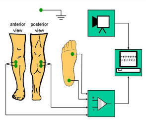
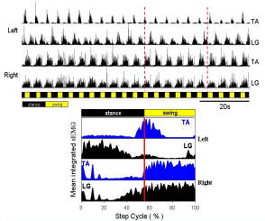
EMG recordings from spinal cord injury patients
The EMG data are obtained using differential surface recordings from the lateral
and medial gastrocnemius (LG and MG) and tibialis anterior (TA) muscles in spinal cord
injury patients during body-weight supported treadmill locomotion using driven robotic
gait orthosis (Etlin et al. 2007). Recordings are synchronized with an analog video
camera using a time code generator controlled by video image acquisition software.
Genetic targeting of specific populations of spinal interneurons
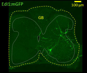
Interneuron-specific enhancer elements for genes expressed in known types of
dorsal interneurons were identified by Prof. Klar's group in our department (see collaborations).
These enhancer elements are cloned upstream of Cre recombinase, injected to the neural tube of the developing (E3)
chick embryo with a conditional GFP or GCaMP3 plasmids, and electroporated in ovo into specific populations
of dorsal interneurons. Ten to 12 days later (E13-15), the spinal cord is isolated and the segmental and
spatial distribution, axonal trajectories, and connectomes of the genetically identified interneurons are
determined. The activity patterns of known sub-types of interneurons targeted with the genetically encoded
calcium sensor GCaMP3 are imaged and the correlation between the optical and the concurrently recorded motor output
is examined (Etlin et al. 2011, Hadas et al. 2011).


Data acquisition and analyses
Data are continuously recorded, digitized using an A/D converter and
saved on the computer for subsequent off-line analyses. Wavelet and
Wavelet coherence analyses are performed to detect rhythmicity, to
determine the phase/power between time series variables in
time/frequency domain
(Mor and Lev-Tov, 2007,
Etlin et al. 2010, Mor et al. 2011).
Descriptive statistical methods of circular distributions and hypothesis testing of these
distributions are also used to describe and test the phase relation
of selected regions of the wavelet spectra
(Mor and Lev-Tov, 2007,
Etlin et al. 2010).
