Coordinated activation of rhythmogenic networks in the spinal cord
The hindlimb locomotor CPGs of the rodents are distributed along the thoraco-lumbar
segments of the spinal cord. We found that an additional rhythmogenic network resides
in the sacrocaudal segments of the spinal cord. This network controls the axial and tail musculature of rodents.
It is tightly coupled to the locomotor pattern generators to ensure body stabilization during movements.
The rostrocaudal coupling between the locomotor and body-stabilizing networks is mediated by propriospinal/descending neurons,
the axons of which travel through the lateral (VLF, LF and DLF) and ventral (VF) white matter funiculi.
Caudo-rostral coupling between the two networks was found during noradrenergic activation of the sacral segments.
This coupling accounts for an increase in the excitability of rostral lumbar motoneurons and the resultant increased
probability for their recruitment

Interactions between afferent pathways and pattern generators
Afferent input has been shown to play a major role in our capacity to reactivate spinal circuits that generate
coordinated rhythmic flexion and extension of the limb muscles in spinal cord injury patients. Our studies revealed
that stimulation of sacrocaudal afferents (SCA) is a potent means for activating the locomotor central pattern generators (CPGs)
in rodents spinal cords that lack the descending control from the brain. These studies showed that capacity of SCA to
induce the locomotor rhythm depends on activation of sacrocaudal neurons the crossed and uncrossed projections of which,
ascend through the ventral and lateral white matter funiculi (VF,VLF, LF, and DLF) to the limb innervating segments of the cord.
The project examines the axonal projections, spatial distribution, organization and physiological properties of these
sacrocaudal neurons and evaluates their role in the generation of afferent induced rhythmicity in the spinal cord.
The studies are performed in collaboration with
Dr. L. Anglister and E.
Finkel, from our department and with
Drs. M.J. O'Donovan and G. Z Mentis
from the NINDS, NIH, USA.
(Anglister et al. 2008,
Lev-Tov and O'Donovan 2009,
Lev-Tov et al. 2010,
Etlin et. al. 2010)

The role of genetically characterized populations of dorsal interneurons in sensorimotor integration
The coordination of locomotion by the central nervous system depends on proper wiring within and
between interneuron populations that make up the complex spinal neuronal networks and on the processing
of the multimodal sensory input delivered via afferent neurons to these networks. Genetic studies revealed
that at least 8 different groups of interneurons develop from the dorsal aspect of the embryonic neural tube.
These dorsal interneurons (dIN) mature to form the various types of tract- and propriospinal neurons that are
critical for sensory-motor integration in the central nervous system. The goal of the project is to determine
the functional connectivity of known types of dINs and examine their role in the neural control of movements.
We use gene targeting and optogenetics as means for tracking and manipulating, activating and silencing identified
subpopulations of spinal interneurons that are required for activation of spinal neuronal networks and for sensorimotor integration.
This project is done in collaboration with Prof. A. Klar and Y. Hadas in our department
(Hadas et al. 2011, Etlin et al, 2011).
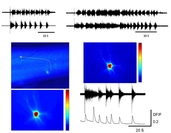
Statistical analyses of non-stationary physiological and pathophysiological activity of the nervous system
The detection of firing patterns embedded within a complex nonstationary
signal and noisy background activity is crucial for the interpretation
of electrophysiological recordings from the brain, spinal cord, and the
neuromuscular system under normal and pathological conditions. In a recent work we have used
Wavelet Transformation
(WT) for the analyses
of non-stationary patterns produced by the mammalian spinal cord preparations
(Mor
and Lev-Tov, 2007, Etlin et al, 2010).
The WT
WT/coherence algorithms enable us to characterize the dynamic profile of the rhythmic signals,
to assess the linear relation between spectra of any given pair of variables, and to uncover and
extract hidden components of the rhythm in the time/frequency domain. These algorithms are integral
parts of "Spinalcore" a menu-driven statistical
package we developed. The computational efficiency of "Spinalcore"
is now improved by graphical processing units (GPUs) based parallel computing (Mor et al. 2011).
The use of "Spinalcore" is expanded these days for
the detection and analysis of pathological and pathophysiological activity of the central nervous system.

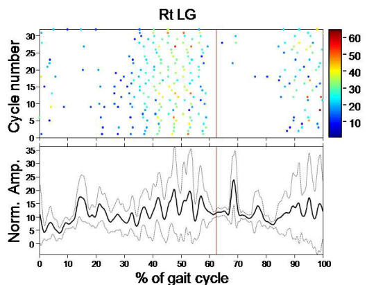
Studies of spinal cord injury patients
The capabilities of the WT time series modules of "Spinalcore" to decompose
non-stationary signals in the time/frequency domain are exploited in this project
for studying the pathophysiology of different forms of spastic activity in spinal cord
injury patients during body weight supported (BWS) treadmill locomotion using driven
robotic gait orthosis. The studies are aimed at reducing the occurrence of the spastic
episodes and enhancing the motor capacity of the patients, are performed in collaboration with
Dr. G.
Zeilig and B. Shamir from the Department of Rehabilitation Neurology
at the Sheba Medical Center, Tel Hashomer.